Red Green Colorblind Test
The Cambridge Color Test for ViSaGe provides a rapid means of screening subjects for colour vision deficiencies.
- The test shows you various Ishihara plates, each displaying numbers that you need to identify correctly to pass this test.
- Remove any glasses with colored lenses. This test requires you to use your naked eye for accurate results.
- Increase your screen brightness to a high level. Dim lighting can affect your ability to see colors accurately.
- For better results, take this test on a larger screen, like a tablet or laptop.
CAMBRIDGE COLOR TEST
The Cambridge color blindness test for ViSaGe gives a rapid means of screening subjects for color vision deficiencies. It also examines the changes in color discrimination that occur as a result of congenital or acquired conditions. ( Research Paper)
INVESTIGATE THE LIMITS OF COLOR DISCRIMINATION
This test allows the investigator to monitor quantitatively over time the progress or remission of the disease. Many drugs addicted people affected with color vision. The pharmacologist will find the test well fitted to monitoring the short-term or long-term side-effects of the blindness.
The test determines discrimination ellipses in color-deficient subjects by probing chromatic sensitivity along the color confusion lines. Generated Ellipses measured in individuals with even slightly anomalous color vision are characteristically orientated and enlarged.
EASY TO USE
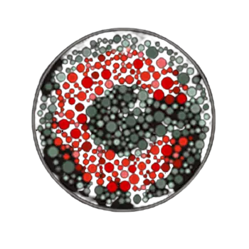
The Cambridge test is easy to use for people with color blindness people or without blindness people. This test uses a familiar Landolt C stimulus, defined by the two test colors that are to be discriminated, on an achromatic background.
The test uses the fixed concept of introducing spatial and luminance noise into the stimulus, composed of grouped circles randomly varying in diameter and having no spatial structure.
RESULTS
Results are saved in ASCII (American Standard Code For Information Interchange) format and presented graphically as discrimination ellipses in CIELUV or CIE(x,y) color space.
The results are typical of a subject with normal color vision; in deficient subjects, the discrimination ellipses are significantly extended in the protan, deutan or Tritan chromaticity directions.


